


Table of Contents
Overview
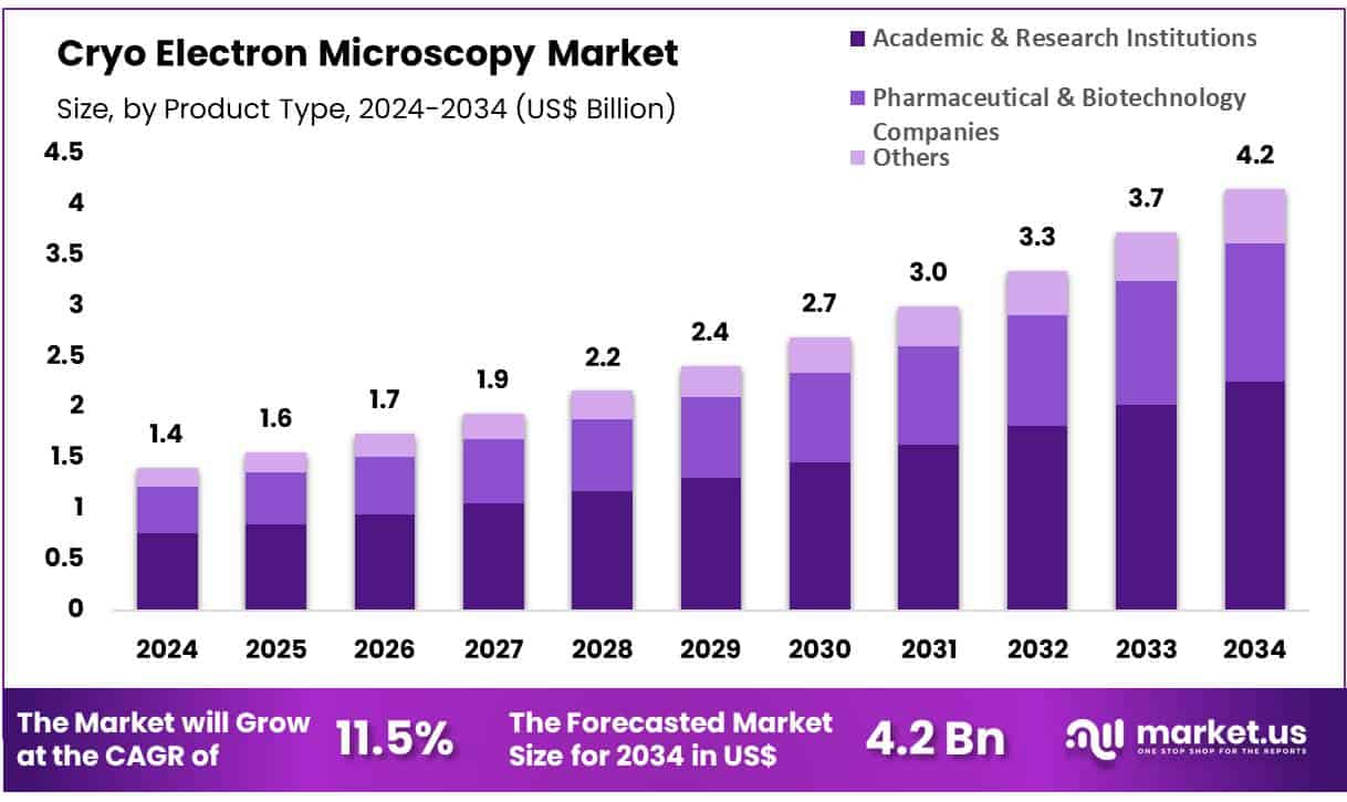
New York, NY – June 02, 2025 – Global Cryo Electron Microscopy Market size is expected to be worth around US$ 4.2 billion by 2034 from US$ 1.4 billion in 2024, growing at a CAGR of 11.5% during the forecast period 2025 to 2034.
Cryo-electron microscopy (Cryo-EM) is revolutionizing the field of structural biology by enabling scientists to visualize biomolecules at near-atomic resolution without the need for crystallization. This advanced imaging technique involves rapidly freezing biological samples at cryogenic temperatures, thereby preserving their native structures. The samples are then examined using an electron microscope, producing high-resolution 3D images that reveal intricate molecular details.
Unlike traditional methods such as X-ray crystallography, Cryo-EM allows the observation of complex and dynamic biomolecular assemblies in their natural environments. It has become particularly valuable in studying large protein complexes, viruses, and cellular organelles. The ability to capture multiple conformational states of molecules has led to deeper insights into biological processes and mechanisms of disease.
In recent years, Cryo-EM has gained widespread recognition for its role in drug discovery, vaccine development, and structural virology. The 2017 Nobel Prize in Chemistry, awarded for advancements in Cryo-EM, marked a pivotal moment for the technique. Its adoption across pharmaceutical and research institutions has increased significantly, supported by ongoing improvements in detector technologies, image processing algorithms, and automation.
As global investment in Cryo-EM continues to rise, it is positioned as a cornerstone technology in the next generation of biomedical research, offering precise molecular insights essential for innovative therapeutic development.
Key Takeaways
- In 2023, the global Cryo-Electron Microscopy (Cryo-EM) market generated revenue of USD 4.0 billion and is projected to reach USD 4.2 billion by 2033, expanding at a CAGR of 11.5% during the forecast period.
- Based on product type, the market is segmented into scanning electron microscopes, transmission electron microscopes, and accessories & software. Among these, transmission electron microscopes dominated the segment with a market share of 47.8% in 2023.
- By application, Cryo-EM is utilized across various domains including biological science, nanotechnology, life sciences, material science, medical, semiconductor, and others. The biological science segment held the largest share, accounting for 38.7% of the market in 2023.
- Regarding end users, the market is categorized into academic & research institutions, pharmaceutical & biotechnology companies, and others. The academic & research institutions segment led the market with a dominant 54.3% revenue share in 2023.
- Regionally, North America emerged as the leading market, capturing a 39.6% share of global revenue in 2023, supported by advanced infrastructure and high adoption of Cryo-EM in scientific research.
Segmentation Analysis
- Product Type Analysis: In 2023, transmission electron microscopes (TEM) led the product segment with a 47.8% market share, driven by rising demand for high-resolution imaging in biological and material sciences. TEMs offer atomic-level visualization, essential for structural biology and nanotechnology research. Their ability to deliver precise, artifact-free images supports advanced studies of proteins and viruses. Ongoing innovations in cryo-EM techniques are further expected to boost the adoption of TEM systems across scientific and industrial research applications.
- Application Analysis: Biological sciences accounted for 38.7% of the cryo-EM market in 2023, reflecting increased use in studying biomolecular structures. Cryo-EM enables high-resolution imaging of macromolecular complexes and viruses in their natural states without crystallization. This has become critical for drug development, disease mechanism research, and therapeutic discovery. As life sciences and molecular medicine demand more precise structural insights, the biological sciences segment is expected to maintain strong growth throughout the forecast period.
- End-User Analysis: Academic and research institutions led the end-user segment with a 54.3% revenue share in 2023, fueled by increased investment in advanced imaging technologies. These institutions are major adopters of cryo-EM for high-resolution biomolecular analysis and structural biology research. Expanding funding for scientific innovation and a growing number of medical and pharmaceutical studies are anticipated to further elevate cryo-EM usage in academic settings, reinforcing their dominant role in market expansion.
Market Segments
Product Type
- Scanning Electron Microscopes
- Transmission Electron Microscopes
- Accessories & Software
Application
- Biological Science
- Nanotechnology
- Life sciences
- Material Science
- Medical
- Semiconductor
- Others
End-User
- Academic & Research Institutions
- Pharmaceutical & Biotechnology Companies
- Others
Regional Analysis
North America led the global cryo-electron microscopy (Cryo-EM) market in 2023, accounting for a 39.6% revenue share. This dominance is attributed to strong investments in advanced research infrastructure and the widespread adoption of high-resolution imaging in structural biology and pharmaceutical research. The launch of innovative technologies, such as Leica Microsystems’ Coral Cryo-electron workflow in 2022, enhanced imaging precision and workflow efficiency, supporting scientific advancements. The region also benefits from a robust ecosystem of academic institutions, biotech firms, and industry collaborations, supported by substantial government and private funding.
In contrast, the Asia Pacific region is projected to register the highest compound annual growth rate (CAGR) during the forecast period. Growth is driven by increasing investments in life sciences research, government R&D initiatives, and national programs like China’s “Made in China 2025.” Expanding biotech hubs, academic collaborations, and rising demand for structural biology tools are supporting market expansion. Additionally, local manufacturers are expected to offer cost-effective cryo-EM systems, boosting accessibility. Enhanced training programs and increased institutional funding are also contributing to the region’s growing capabilities in high-end scientific imaging.
Emerging Trends
- Advancements in Instrumentation and Automation: The capabilities of cryo-electron microscopy (cryo-EM) have been significantly enhanced by improvements in detector technology and automated workflows. High-frame-rate direct electron detectors now allow images to be captured with greater sensitivity and less noise, which has led to faster data collection and higher resolution maps. Automated sample loading and grid screening systems have reduced manual intervention, decreasing the time needed for data acquisition and processing.
- Expansion of Cryo-Electron Tomography (Cryo-ET): Cryo-ET methods have been increasingly used to visualize cellular machinery in its native context. Recent developments have enabled the imaging of rare assemblies and difficult-to-access cellular regions, such as membrane-bound complexes within intact cells. The implementation of focused ion beam milling and improved software for tilt-series alignment has allowed reconstructions of subcellular volumes at resolutions sufficient to discern macromolecular arrangements.
- Integration of Machine Learning and AI: Artificial intelligence (AI) and deep learning approaches are being integrated into cryo-EM data processing pipelines to speed up tasks such as particle picking and de novo model building. New algorithms can automatically identify protein densities, reducing manual annotation time by up to 70%. AI-based tools are also being used to enhance map sharpening and to guide model reconstruction, which has led to faster atomic model generation from density maps.
- Growth of Public Data Repositories: The volume of cryo-EM structures deposited in public databases has been rising rapidly. As of 28 May 2025, the Electron Microscopy Data Bank (EMDB) contained 46,065 entries, up from roughly 30,000 entries in 2021. Similarly, 24,084 three-dimensional electron microscopy (3DEM) structures were released in the Protein Data Bank (PDB) in 2024, compared with 18,292 in 2023. This trend reflects both an increase in global cryo-EM capacity and the standardization of data-sharing practices.
Use Cases
- Viral Structure Determination for Vaccine and Antiviral Design: Cryo-EM has been instrumental in revealing the structures of viral surface proteins at near-atomic resolution. During the COVID-19 pandemic, cryo-EM maps of the SARS-CoV-2 spike trimer were generated at \~3.5 Å resolution, enabling rapid design of stabilized spike immunogens. The Dubochet Center for Imaging (DCI) resolved the Omicron variant’s spike structure within weeks of its identification, aiding in updated vaccine development. In 2024 alone, over 5,000 viral structures were deposited to EMDB, underscoring cryo-EM’s role in expedited viral research.
- Membrane Protein Structural Proteomics: Recent studies have demonstrated that small (< 100 kDa) and low-abundance (< 10 %) membrane proteins can be resolved within heterogeneous samples. For example, a membrane transporter complex was solved at 3.2 Å resolution despite accounting for less than 5 % of the total protein content in the sample. This has expanded the ability to study drug targets and transporters that were previously intractable by X-ray crystallography or NMR. In 2024, over 1,000 membrane protein structures were released via cryo-EM, up from just 250 in 2010, illustrating a fourfold increase in such entries over 15 years.
- Amyloid and Neurodegenerative Disease Research: Cryo-EM has permitted the visualization of amyloid filaments implicated in diseases like Alzheimer’s, Parkinson’s, and prion disorders at atomic resolution. High-resolution maps (2.8–3.2 Å) of Aβ and tau filaments were obtained, providing insights into filament polymorphisms and potential small-molecule binding sites. In studies published in 2023, over 20 unique amyloid fibril structures were reported, enabling structure-based design of aggregation inhibitors.
- Drug Discovery and Rational Design of Therapeutics: Pharmaceutical research has leveraged cryo-EM to determine the binding modes of small molecules and biologics to their protein targets. By mapping ligand–protein interactions at \~2.5 Å resolution, medicinal chemists have optimized inhibitors against challenging targets such as ion channels and GPCR complexes. In 2024, approximately 250 cryo-EM structures of drug–target complexes were published, accelerating lead optimization cycles by up to 40 % compared to traditional methods.
- In Situ Structural Biology via Cryo-Correlative Light and Electron Microscopy (Cryo-CLEM): The combination of fluorescence microscopy with cryo-EM (cryo-CLEM) has enabled the study of macromolecular complexes within intact cells at nanometer resolution. For example, analyses of mitochondria-associated ribosomes in neuron cultures achieved \~10 Å resolution within cryo-preserved cells, revealing conformational changes during translation. In 2024, over 150 in situ structures were reported, marking a tenfold increase since 2018.
Conclusion
Cryo-electron microscopy (Cryo-EM) is transforming biomedical research by providing near-atomic resolution imaging of complex biological structures without the need for crystallization. Its applications span from vaccine development and drug discovery to neurodegenerative disease research and structural virology.
The technology’s rapid adoption is driven by innovations in automation, AI integration, and increased public data sharing. As academic institutions, pharmaceutical companies, and research organizations continue to invest in cryo-EM systems, the technique is poised to remain a critical tool in structural biology, accelerating scientific breakthroughs and enabling the design of targeted therapies with unprecedented molecular precision.
Discuss your needs with our analyst
Please share your requirements with more details so our analyst can check if they can solve your problem(s)